Project status: OPen – looking for partnership
3D printing, or rapid prototyping, is a rising and wide-spreading technique that can be successfully used to create anatomically accurate models from medical imaging studies. Applications in biology and medicine range from diagnostic to educational purposes, as well as for surgical planning or prosthetics design.
To date, desktop-sized printers with limited costs are an interesting reality, whereas commercial, high-end printer can deliver hi-resolution color models using multiple materials.
The term “rapid”, however, should not be taken literaly; although the printing phase itself may be relatively short, creating a highly complex 3D mesh model may be a time consuming task, especially when using a set of in-vivo images.
Data selection, theshold adjustment, segmentation and surface extraction phases requires lots of resources. Moreover, creating a model requires many other factors to be considered, like file format and size, number of polygons, errors in model, minimum wall thickness, total printing costs, printing times and so on.
Many software available for medical image viewing (like the wonderful Slicer) includes tools and algorithms to create a three dimensional, polygonal model from a set of images.
Marching cube algorithm (REF) is one of the most diffuse techinique for reacreating 3D isosurfaces (isocontour) from a 2d-stack of images. User defines a threshold value (eg. in Hounsfield units for TC images) and the algorithm will automatically process the images by extracting a polygonal mesh to be exported in WRL or similar file format to perform further editing, 3D printing or rendering.
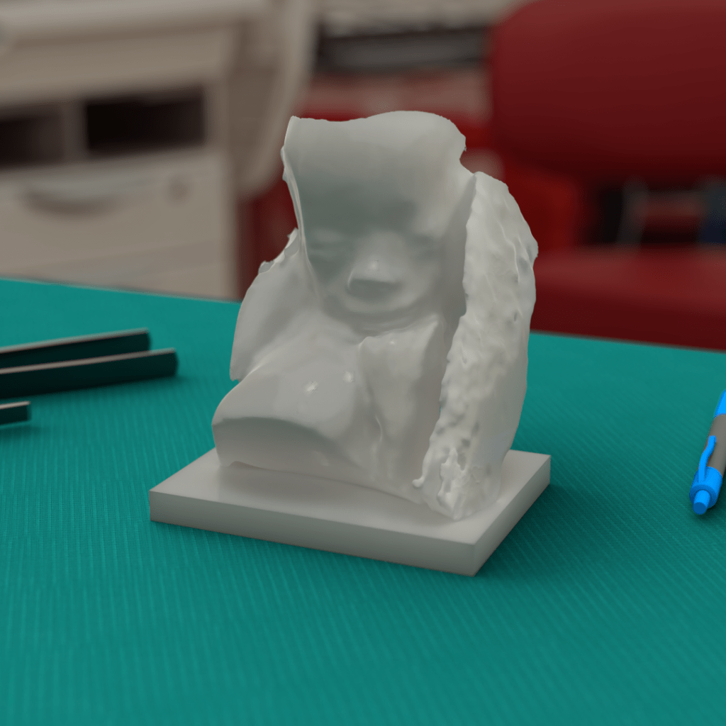
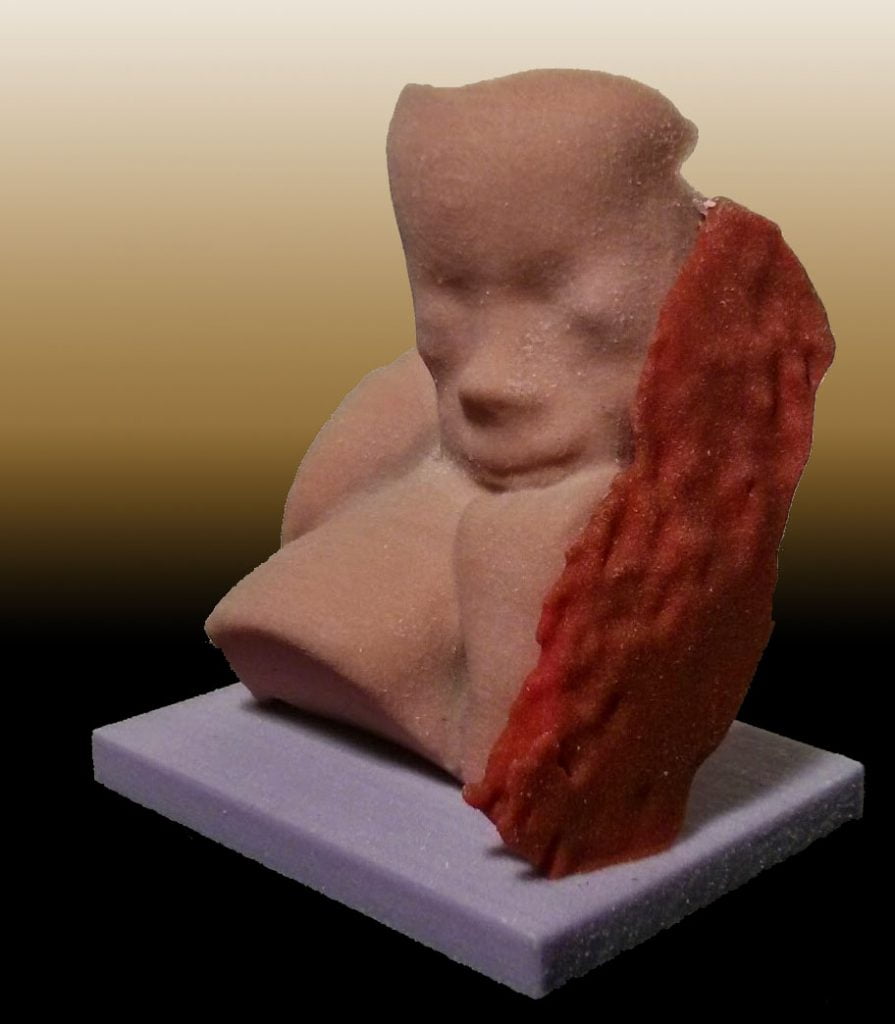
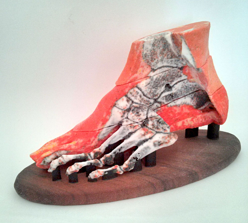
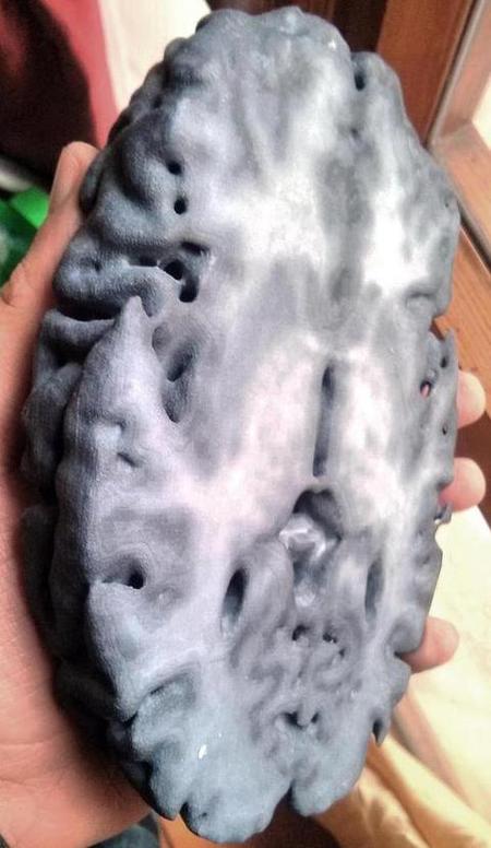
In most cases, however, these model are monochromatic and lack color and texture information: being the results of a marching cube algorithm, they are -by definition- an iso-surface and don’t retain any color information from the original scan series.
CRAFTER is a custom software (written in Processing) that allows the creation of 3D (WRL) models of a set of contiguous medical images. The name “CRAFTER” is an homage to Minecraft, a voxel-based game that inspired this method.
Medical images, once exported in a standard .jpg format, are imported in CRAFTER. The software provides a custom color palette (balck-white gradient, black-red, and so on) and then creates a ready-to print WRL file.
WRL was chosen because it has a quite simple standard format. This can be easily converted in any other format, like STL or OBJ.
CRAFTER is available as a pre-alpha release. Some things should still be impemented:
- GUI
- Some debugging (I’m a physician, not a programmer…so the code is quite ugly)
- Re-wirte the code in C++ or similar
- STL file export
- Create a github repository
- Submit a sci-paper
I’m looking for programmers and researchers willing to create a final version and/or write down an almost complete paper.